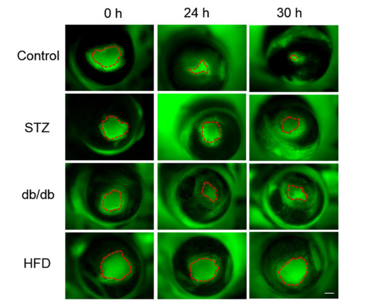
People with diabetes often suffer from wounds that are slow to heal and can lead to ulcers, gangrene and amputation. New research from an international group led by Min Zhao, professor of ophthalmology and of dermatology at the University of California, Davis, shows that, in animal models of diabetes, slow healing is associated with weaker electrical currents in wounds. The results could ultimately open up new approaches for managing diabetic patients.
[adsense:336x280:8701650588]
"This is the first demonstration, in diabetic wounds or any chronic wounds, that the naturally occurring electrical signal is impaired and correlated with delayed healing," Zhao said. "Correcting this defect offers a totally new approach for chronic and nonhealing wounds in diabetes."
It has been estimated that as much as $25 billion a year is spent on treating chronic ulcers and wounds related to diabetes, Zhao said.
Electric fields and wound healing
Electric fields are associated with living tissue. Previous work by Zhao and Brian Reid, project scientist at the UC Davis Department of Dermatology, showed that electric fields are associated with healing damage to the cornea, the transparent outer layer of the eye.
In the new work, published June 10 in the journal Scientific Reports, Zhao, Reid and colleagues used a highly sensitive probe to measure electrical fields in the corneas of isolated eyes from three different lab mouse models with different types of diabetes: genetic, drug-induced and in mice fed a high-fat diet.
In a healthy eye, there is an electrical potential across the thickness of the cornea. Removing a small piece of cornea collapses this potential and creates electric currents, especially at the edges of the wound. Cells migrate along the electric currents, closing the scratch wound in about 48 hours.
The researchers found that these electric currents were much weaker in eyes from all three strains of diabetic mice than in healthy mice. Delayed wound healing was correlated with weaker electric currents.

Caption: UC Davis researchers measured electric fields and wound healing in eyes from three different models of diabetes. Left to right, green fluorescence shows damaged area shrinking over time. Top row, eyes from normal mice. Other rows are eyes from three different mouse models of diabetes.
Credit: Min Zhao and Brian Reid, UC Davis
"We saw similar results with all three models," Reid said.
The researchers also found that human corneal cells exposed to high levels of glucose showed less response to an electric field. Diabetics have high levels of glucose in their tears, Reid noted.
[adsense:468x15:2204050025]
Unique facility
The UC Davis bioelectricity laboratory is one of a very few able to make such sensitive measurements of electric fields in living tissue.
"We might be the only lab in the country that is able to do this," Reid said. They are collaborating with a number of laboratories worldwide and across the country, as well as several other UC Davis departments.
Sources: University of California - Davis


