About Authors:
Aditi Singhal*, Dharmendra Kumar, Mayank Bansal
Department of Pharmaceutical Technology,
Meerut Institute of Engineering and Technology,
Baghpat bypass crossing, NH-58, Delhi-Roorkee Highway, Meerut-250005, U.P., India.
*singhal28aditi@gmail.com
Abstract:
It is the time for herbal cosmetics as it is the time for looking young and smarter. They are also being widely used for improving several skin related problems. Herbal cosmetics are also called as natural cosmetics. Skin care cosmetics are being used for several functions like anti-oxidants, anti-inflammatory, anti-microbial, anti-septic etc. vitamins are also added in skin care cosmetic formulation. Pigmentation is the reason for various skin related diseases like skin darkens, aging, acne, wrinkles etc. Anti-oxidant property, anti-microbial activity, skin moisture content activity, mushroom tyrosinase inhibitory assay etc are various methods for the evaluation of skin care cosmetics. Natural Cosmaceuticals does not have any side effects to the skin, easy to handle, easy to store, easy to use and offers various beneficial effects to the skin.
REFERENCE ID: PHARMATUTOR-ART-1853
Introduction
1. Anatomy and Physiology: [1,2,3,4]
Skin is the body’s outer covering layer which helps to protect us from heat, light, microbial infection and injury. It is the body’s largest organ, mainly regulates body temperature and stores vitamin D which is essential for human body, fat as well as water. Skin is composed of three layers, which are:
1.1.Cellular epidermis.
1.2.Dermis.
1.3.Subcutaneous Tissues.
pH value of skin varies from 4.0 to 5.6. Generally, it is considered as the smooth layer but because of aging, sunrays, dust, microbial infection, cold and heat it becomes rougher and thicker. Now a days, wrinkles and skin darkens are also considered as a major problem. Skin contains mainly two types of essential fatty acids, linoleic acid and arachidonic acid, having a vital role in regulating barrier functions. Two types of skin are present in humans, hairy and non-hairy. Hair follicles and sebaceous glands present in hairy skin and non-hairy skin consists only of sebaceous glands. Non-hairy skin is present in palms and soles.
1.1. Epidermis:[3,5,6]The outer layer of skin works as a protective sheath against environmental influences like sunrays, dust, heat, microbial infection etc. Epidermis comprises of five separate layers like;
a. Horny layer (Stratum corneum).
b. Stratum lucidum.
c. Stratum granulosum (Granular layer).
d. Stratum spinosum (Prickly cell layer).
e. Stratum germinativum (Basal layer and dermoepidermal junction).
1.2. Dermis: [3]
Dermis is the middle layer of skin which gives elasticity, moisture and firmness to the skin. Blood vessels, nerve cells, sebaceous glands, sweat glands are present in dermis layer. Dermis is having 0.2 to 0.3cm thickness. It also contains dense network of structural protein fibers that is collagen, reticulum and elastin.
1.3. Subcutaneous tissues: [3]
Subcutaneous layer consists of a sheet of fat areolar tissue. This type of layer is quite elastic and having the presence of large arteries and veins in the superficial region. It mainly synthesizes and stores high energy chemicals.
2. Skin disorders: [7,8]
2.1. Pigmenting disorders- These are also called as pigmentation disorders and are due to excessive or reductive production of melanin. Due to this disorder, skin becomes darkens, blotchy (discolored) or lighter.
2.1.1. Types of Skin Pigmentation:
2.1.1.1. Hypo-pigmentation- It is caused by lesser amount of melanin. It can also occur in caucasions. The common disorders due to hypo-pigmentation are-
2.1.1.1.1 Vitiligo-It is an auto-immune disorder which causes white spots on the skin. Now a day’s 2% of the population is affected by vitiligo. Addison’s disease and hyperthyroidism are common with vitiligo.
2.1.1.1.2 Albinism- It occurs due to complete absence of pigment. Patient suffering from this disorder have pale skin and light yellow eyes. Skin cancer is common with albinism.
2.1.1.2. Hyper-pigmentation- It is caused by excessive production of melanin in the skin. The major factors which lead to hyper-pigmentation are excessive amount of sunbathing and malnutrition. Drugs like antibiotics, anti-malarial, anti-arrhythmic can also lead to hyper-pigmentation. The common hyper-pigmentation disorders are lamellar ichthyosis (fish scale disease), lichen simplex and melasma.
2.2. Sebaceous and Sweat glands disorders- The common disorders are acne, pimples, blackheads, prickly heat. This disorder mainly occurs in neck and large area of skin.
2.3. Skin scaling disorders- The commondisorders are dandruff and psoriasis.
2.3.1.Psoriasis- It is the formation of scaly red patches on skin.
2.3.2. Dandruff- It can be caused by microbial infection or immunological disorders. It is present on the stratum corneum.
2.3.3.Effects of aging on skin- Due to aging skin become darker, dull, damage and lower melanin level. Acne and wrinkle are very common examples.
3. Skin problems treated by herbal and Synthetic (man-made) cosmetics:[9]
Skin problems are generally treated by the help of herbal and synthetic cosmetics. Cosmetics are used to reduce/lower the wrinkles, acne, darkness and controls oil secretion. Various skin problems are occurred by the excessive amount of sunrays bathing, aging and wrinkles. These problems are treated by using cosmetics which are having antioxidant, anti-inflammatory, antibacterial and antiseptic properties. Herbal and Synthetic preparations are available in market in the form of creams/gels. Massage creams, vanishing creams and face wash gels are also used to treat various skin problems. To protect from U.V. radiations various sun protecting factors like SPF 15, SPF 30, SPF 40, SPF 50 are used but SPF 40 and SPF 50 are best suited to protect skin from U.V. radiations. In herbal cosmetics, natural substances like aloe-vera, cucumber, turmeric, green tea, rose etc. are used to treat all types of skin problems. Similarly synthetic cosmetics like pearl powder, Sebum REG, BHT (butylated hydroxyl toluene), Beeswax, Candelilla wax, Lanolin alcohol, Ozokerite wax, Ethylene diamine tetra acetate (EDTA), Triethanolamine etc are used to treat skin problem. Skin diseases are very common symptoms of lupus. Almost 80% of the lupus suffering patients are having several skin related problems/diseases. Mainly three types of lupus are as follows; [10]
3.1. Acute cutaneous lupus erythematosus (ACLE).
3.2. Subacute cutaneous lupus erythematous (SCLE).
3.3. Discoid lupus erythematous (DLE) or Chronic cutaneous lupus erythematous (CCLE).
3.1.Acute cutaneous lupus erythematosus (ACLE) – It is also known as ‘Malar rash’ which involves the upper cheeks, and is also known as ‘Butterfly rash’ which refers unique butterfly like shape.
3.1.1. Characteristics of ACLE-
a. ACLE is having a symmetrical appearance which covers both cheeks and the nose bridge.
b. Neck and forehead are also affected in many cases.
c. Skin becomes red and swollen similarly like sunburn.
3.1.2. Causes and long term effects-
ACLE is highly photosensitive. ACLE lasts for several weeks or days. It may cause skin rashes, but if the rashes will clear there will be no permanent effects.
3.2.Subacute cutaneous lupus erythematosus (SCLE) –
3.2.1. Characteristics of SCLE-
a. Ring shaped or dish- shaped patches will occur.
b. Sun exposure and another form of U.V. light may cause SCLE.
3.2.2. Causes and long term effects-
SCLE is also photosensitive; it is started by U.V. light. Skin becomes darkening due to this disorder. It can also occur due to side effects of medication.
3.3.Discoid lupus erythematosus (DLE) – It is also called as Chronic cutaneous lupus erythematous (CCLE).
3.3.1. Types of Discoid lupus erythematosus (DLE) -
a. Localized DLE- The lesions are located on the scalp and face.
b. Generalized DLE- Thelesions are present on any portion of the body.
3.3.2. Characteristics of DLE-
a. It may only affect the skin.
b. Disk or coin shaped lesions occur.
c. Painless.
d. Usually present on scalp and face.
3.3.3. Causes and long term effects-
DLE is more photosensitive then SCLE. It may damage hair follicles which lead to permanent hair loss. Several environm1111ental factors and different types of medications may used to stimulate the symptoms of DLE.
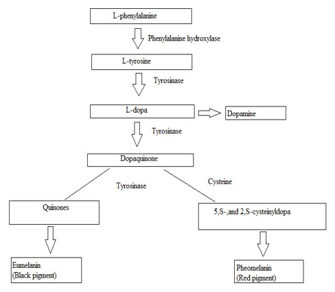
4. Melanin Pigment- Melanin is the end-product of various multiple transformations of L-tyrosine. Melanin is biosynthesized by;
a. Initiated by the hydroxylation of L-phenylalanine to L-tyrosine.
b. Oxidation of L-DOPA to dopaquinone- is similar for both eumelanogenesis and pheomelanogenic pathway. [11]
Eumelanogenesis involves the procedure of dopaquinone to leukodopachrome. Pheomelanogenesis may also starts with dopaquinone, conjugate with cysteine or glutathione to yield cysteinyldopa and glutathionyldopa for transformation into pheomelanin. Melanin pigments having their common arrangements linked by carbon-carbon bonds, but having different chemical composition, physical as well as chemical properties. Eumelanine is behaving like polyanions, having the capability of reversibly bind anions, cations and polyamines. Electron paramagnetic resonance (EPR) spectrum of eumelanin yields to slightly asymmetric singlet which produces a free radical signal. Pheomelanin is the back bone of benzothiazine units which exhibits a yellow to reddish brown color and is soluble in alkali. Pheomelanin is highly protein bound which indicates that it acts like a chromo protein [11,12,13], it can also act as a binding agent for chemicals and drugs.[14] Adult female epidermis is less melanized in comparison to males due to gender specific effect.[15] Function of melanin in human skin appears by the attenuation of U.V. penetration to the blood in dermal vessels.[16].

Fig 1: Melanin Formation
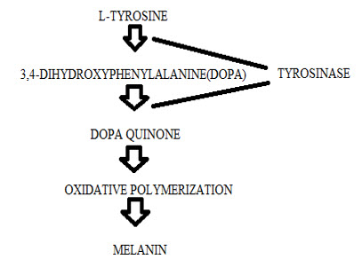
5. Melanogenesis Pathway- Melanocytes are usually 7µm in length.Melanin is the only factor responsible for skin coloration. Melanin is produced in the melanocytes, where melanocytes stored in the melanosomes after these melanocytes transferred melanosomes into keratinocytes. Melanocytes are stored in the epidermal-dermal junction. They extend the intracellular nitric acid (NO) production which initiates signal transduction cascades for the production of Melanogenesis by the help of tyrosinase enzyme. [17].

Fig.2: Melanogenesis Pathway
5.1 The process of Melanogenesis can be understood in following three steps-Melanogenesis process is done by three steps such as; [18]
a. Production of Cysteinyldopa- The process occurs with the addition of cysteine to dopaquinone.
b. Oxidation of Cysteinyldopa- This process gives the pheomelanin.
c. Production of eumelanin- which starts after the reduction of Cysteinyldopa.
5.2 Mechanism of regulation of Melanogenesis: [19-35]
Melanogenesis is regulated by two methods which are;
5.2.1. Transcriptional Regulation- Tyrosinase genes generally contains at least one major and three minor transcription sites.[19] TATA and CAAT boxes comes under promoter region, many potentional micropthalmia-associated transcription factor (MITF) binding sites including E-boxes, M-boxes as well as tyrosinase distal element (TDE), five AP-1 and two AP-2 binding sites are used for phorbol ester-inducible enhancer-binding protein AP-1 (Activator protein-1), UV responsive elements (URE), thyroid and retinoic acid like responsive elements (TRE and RER), tyrosinase element-1 (TE-1) [20-30]. Transcriptional regulation of Melanogenesis related genes is having an important role by the number of class III POU binding protein Brn-2/N-Oct3 [31,32]. Negative transcriptional regulation process is mediated by brachyury-related transcription factor (TBX 2) [33].
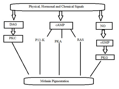
5.2.2. Intracellular signal transduction regulation- Intracellular signal transduction pathway is used for the positive regulation of Melanogenesis with cAMP used as the critical factor[34,35].

Fig.3: Intracellular Signal Transduction Pathway
Where, DAG- diacylglycerol ; PKC- Protein kinase C ; P13-K- Phosphatidylinositol 3-kinase ; PKA- Protein kinase A ; NO- Nitric acid ; PKG- Protein kinase G.
cAMP regulation mechanism of Melanogenesis including the activities of protein kinase A (PKA), then phosphorylates enzymes, ion channels and various regulatory proteins. Transcriptional activity is regulated by activated PKA (Protein kinase A) which involves phosphorylation of cAMP responsive element binding protein (CREB) and CREB binding protein (CBP).
6. Herbal cosmetics: [36]
Herbal cosmetics have a great role for reducing the skin problems which occurs due to several factors. Herbal cosmetics have no side effects while applying on skin. Generally herbal cosmetics are derived from botanical extracts which are having highly antioxidant property, anti inflammatory property and rich in vitamin C and vitamin K. Herbal cosmetics are mostly used by the following benefits;
a. Safely to the skin.
b. Easily available.
c. Low cost.
d. Having less or no side effects on the skin.
e. Having high concentration of vitamins and minerals which are beneficial for prevention of skin disease.
f. Almost more than half population totally depends on the herbal cosmetics as they are having less or negligible side effects.
7. List of Some Herbal medicines used for treating Skin diseases- [37,38]
Table 1
|
Scientific name |
Family |
Uses |
|
Bauhinia Variegata |
Fabaceae |
Anti-oxidant property |
|
Areca catechu |
Arecaceae |
Anti-oxidant property |
|
Aloe Vera |
Liliaceae |
Anti-dermatitis |
|
Green tea |
Theaceae |
Photo-protective |
|
Ginko bilolsa |
Ginkogaceae |
Skin tonic |
|
Allium sativum |
Alliaceae |
Anti-oxidant |
|
Dancus carota |
Apiaceae |
U.V. protection |
|
Curcuma domestica |
Tricholomataceae |
Skin care |
|
Lentinus edodes |
Zingiberaceae |
Anti-oxidant property |
|
Muntingia calbura (Jamaica cherry) |
Elaeocarpaceae |
Skin lightening and anti- oxidant property |
|
Pluchea indica |
Asteraceae |
Anti-oxidant property |
|
Glycyrrhiza glabra |
Leguminosae |
Skin whitener |
|
Thea viridis |
Theaceae |
Anti-oxidant |
|
Crataevea murula |
Capparidaceae |
Anti-aging |
|
Rosemarinus officinalis |
Lamiaceae |
Anti-aging |
|
Buckwheet seeds |
Polygonaceae |
Anti-wrinkle |
|
Psorolia corlifolia |
Fabaceae |
Pigmenting agent |
8. Synthetic Cosmetics: [39]
Synthetic cosmetics are also used for skin care formulations. Mainly the synthetic cosmetic preparation having a large amount of surfactants used like Span 20, Tween 80, Sodium lauryl sulphate, etc. They are less time consuming, easy to use, easy to carry and easy to store. The various ingredients used in synthetic preparations are as follows;
Table 2
|
Synthetic Agents |
Examples |
|
Anti Aging and Anti Wrinkle agents |
Argireline, Pearl powder, Beta Glucan, Coenzyme Q10, Glycan Booster peptide, Ceramide complex |
|
Soothing and Regenerating agents |
Allantoin, Aloe-Vera gel, Colloidal Oatmeal |
|
Oily Skin Regulators |
Sebum REG |
|
Skin Whitening agents |
Alpha- Arbutin, Skin white BLE, Kojic acid, Skin white MSH, Glycolic acid, Aleosin, Hydroquinone, Salicylic acid |
|
Emollients |
Squaline |
|
Humectants |
Glycerin, Sodium PCA, Caprylyl Gylcol EHG, Algae extract and Hyaluronate gel, Propylne glycol, Hyaluronic acid, Sorbital, Urea |
|
Surfactants |
Alkyl sulphonate, Coco betaine, Coco glucose, Sulfosuccinate, Polyglucose, Polyquaternium-87 |
|
Emulsion stabilizers |
Ethylene diamine tetra acetate (EDTA), Triethanolamine |
|
Anti-oxidants |
BHT(Butylated hydroxyl toulene), propyl gallate, Vitamin E |
|
Thickeners |
Lanolin alcohol, , Cetyl alcohol, Carbomer, Acrylate copolymer |
9. Functional Active Agents in Skin care formulations:
9.1Anti Aging agents-[40] Stress is the major factor for aging. Other factors which cause aging are excess amount of calories present in the body, excessive insulin amount, reduced anti oxidant level, etc. Due to aging the skin becomes less elastic, becomes drier, and wrinkles appears in huge amount. Skin aging problem may breakdown the DNA, Collagen, Elastin, Hyaluronic acid and all other molecules present in the skin. Melanotropin activity, Tyrosinase activity becomes larger due to skin aging problem. Reduction of Vitamin C and Vitamin E are the main factors that causes skin aging problem.Anti-aging cream mainly acts to slow or reduce the signs of aging.
9.2Anti Oxidant agents-[41-43] These agents are used to inhibit the several problems of skin. Reactive Oxygen Species (ROS) is the main factor responsible for several skin problems. Anti oxidants are used to inhibit the ROS production. Anti oxidants works at different levels in oxidative process such as;
a. Free radical scavenging.
b. Lipid peroxyl radicals scavenging.
c. Metal ion binding occurs.
d. Removing oxidatively damaged biomolecules.
Ascorbic acid is the main and effective example of anti oxidant used to control sun burn, cell formation and tumors formation problem. Another example of anti oxidants like Vitamin E, Vitamin C, Lentinus edodes, Pluchea indica inhibits the U.V. penetration power.
9.3. Vitamins- Vitamins are the vital compounds which are very beneficial to the body. Vitamins are used in anti wrinkle creams, anti aging creams, skin whitening creams and in the various products which are very beneficial to the skin. Vitamin C is widely used in preparation of cosmetics as it is having an anti oxidant property [44]. Vitamin E is also having an anti oxidant property and used in cosmetics because of this free radical scavenging property [45,46].In human skin it acts as a predominant lipid soluble anti oxidant in the human stratum corneum. They are also used to protect several components like DNA, proteins, reactive oxygen species (ROS) which are caused by UV radiation and by various air pollutants [47]. Vitamin A (Retinol) is also used in the cosmetics and in dietary nutrient required for the growth of bone and for skin keratosis.
9.4. Emollients-[48] Emollients having a skin softening function. They are used to reduce skin dryness, skin scaling, wrinkles and mild irritant contact dermatitis. Several emollients used for skin nourishing are Almond oil, Caster oil, Coconut oil, Grapeseed oil, Meadowfoam seed oil, Hemp seed oil, Jojoba oil, Rose hip oil, Sesame oil, Squaline, etc.
9.5. Skin lightening actives-[49] Skin lightening active agents used to light the skin color which becomes darker due to excessive amount of sunray bathing. There are a lot of skin lightening formulation present in the market which may contain anti oxidant, anti wrinkle, anti aging properties for the removal of several skin related problems. Skin lightening formulations may also contain natural extracts which influences the melanization process. Skin lightening formulations contains different combinations like Arbutin, Azelaic acid, Kjoic acid, Resveratrol, Liquorice. Trichloroacetic acid is used to remove melanin loaded skin tissues.
10. Testing for Skin care formulations- Patients suffering with these types of lupus disorders have to avoid directly exposure of sunlight or U.V. light by using various sun protecting factors like SPF 30, SPF 40 and SPF 50 etc. various anti-malarial drugs and hydroxychloroquine may be highly effective in treating various skin diseases. Skin care formulations to be used for controlling various skin related problems like skin darkens, aging, black spots, acne and many other problems. Now a day’s both herbal as well as synthetic skin care formulations are more generally used. Herbal cosmetics formulations are used as they are not having any side effect to the skin but they are not easy to use, not easy to handle that’s why synthetic cosmetics are generally used as they are easy to use and easy to handle. Various tests are performed on skin care formulations such as anti- oxidant property test, anti- tyrosinase test etc.
10.1. Anti oxidant property-These agents are used to inhibit several problems occurs with skin. This test is evaluated by several methods like-
10.1.1. DPPH (1, 1-diphenyl-2-picrylhydrazine) radical scavenging activity.
10.1.2. ABTS (2, 2-azino-bis (3-ethylbenzthiazoline-6-sulfonic acid) scavenging activity.
10.1.3. Nitric acid scavenging activity.
10.1.4. Hydrogen peroxide (H2O2) scavenging activity.
10.1.5. Anti oxidant property is also determined by using FRAP (Ferric reducing ability of plasma or plant), Benzie and Strain.
10.1.1. DPPH (1, 1-diphenyl-2-picrylhydrazine) radical scavenging activity-[50] DPPH is commonly used for the anti oxidant property. DPPH method is generally used in a 96 well microtitre plate. When the anti oxidant molecules incubated, they reacts with DPPH (which is in purple color) and forms di-phenyl hydrazine (yellow color). This conversion of DPPH (purple color) to di-phenyl hydrazine (yellow color) measured at 520nm which is used to measure the scavenging potential of the plant extracts added to specified volume of DPPH solution in a microtitre plate. These plates are incubated at 25?C for 10minutes and absorbance is measured at 520nm. DPPH acts as a control solvent and other solvents used with plant extract are used as test solvents.
% inhibition of DPPH radical scavenging activity = [(Absorbance of control-Absorbance of test) / (Absorbance of control)]×100
10.1.2. ABTS(2,2-azino-bis (3-ethylbenzthiazoline-6-sulfonic acid) radical cation decoloration scavenging activity-[51] In this procedure a specified amount of ABTS is dissolved in distilled water and also added potassium persulphate to it. The whole reaction mixture is allowed to stand at room temperature for overnight before use. To various concentrations of extracts, add specified volume of DMSO (di-methyl sulfoxide) and ABTS to make the final volume. Absorbance is measured at 734nm for 20 minutes.
10.1.3. Nitric acid scavenging activity-[52,53] Sodium nitropursside is incubated in phosphate buffer solution (pH 7.4) with different concentrations of test extracts incubated at 25?C for 5 hours. The incubation plates are removed and are diluted with Griess reagent (which is prepared by using equal volume of 1% sulphanilamide in 2% phosphoric acid and 0.1% naphthylethylene dihydrochloride in water). The absorbance is measured at 546nm.
10.1.4. Hydrogen peroxide (H2O2) scavenging activity-[54] Phosphate saline buffer (pH 7.4) is used to prepare hydrogen peroxide solution. Various concentrations of few ml of extracts or standards in ethanol/methanol are added to hydrogen peroxide solution. Absorbance measured at 230nm after 10 minutes. Phosphate buffer without hydrogen peroxide solution (Blank solution) is used to compare it.
10.2. Anti tyrosinase activity-[55] Tyrosinase activity is determined by using dopachrome method where L-DOPA is used as a substrate. The assay method is conducted in a 96-well microtitre plate and absorbance of these plates is measured at 475nm with reference. All samples are dissolved 50% solution of DMSO. Each microtitre plate contains sample and phosphate buffer in the ratio of 1:2 and tyrosinase and L-DOPA in the 1:1 ratio. Results are compared with the 50% DMSO solution in replace of sample.
% tyrosinase inhibition = (Acontrol – Asample) / Acontrol ×100.
10.3. Anti- microbial activity-[56-60]. The anti- microbial activity is performed by the disc-diffusion test by using the specified volume of suspension which is containing few ml of bacteria spread on the nutrient medium. Air dried and powdered fruit materials are extracted in a soxhlet apparatus by using methanol for 72 hours. Extracts are filtered by using Whatmann filter paper and concentrated in vaccum at 40?C by the help of rotary evaporator. Filter paper disc are impregnated with few µg of extract/ disc and placed into agarnutrient. Similarly, negative controls are prepared by using the same solvents used to dissolve plant extracts. The plates are incubated at 35?C for 24 hours.
10.4. Clinical testing- [61] Clinical testing is performed for the determination of results skin of formulations. Clinical testing of skin formulations is generally done on human volunteers or animals who give the exact consent about the formulations. This test is carried out for several weeks or days. The skin formulation (Cosmaceuticals) is applied on all the human volunteers or animals used in study. For evaluation of skin whitening formulations- Chromameter is used for the measurement of skin darkness. Skin darkness is measured which is taken before applying the product and again after few hours. The test carried out for the result of skin formulations.
10.5. Mushroom tyrosinase inhibitory assay-[62] Mushroom tyrosinase inhibitory assay of various plant extracts are determined by spectrophotometric methods. In this method, the plant extracts were dissolved in DMSO solution at a constant concentration then diluted it with different concentrations of DMSO solution. The sample solution is diluted with phosphate buffer pH 6.8 in test tubes. After this add one or two drops of L-Tyrosine solution and finally added mushroom tyrosinase solution. Few microlitres of DMSO and Kojic acid solution are used as blank reference and positive control. The initial absorbance is measured at 490nm. Then incubate it for 20 minutes at 37?C and then the final absorbance is measured at 490nm. The IC50 values are determined the concentration at which the 50% tyrosinase activity inhibited.
% mushroom tyrosinase inhibition = [(A2 – A1) – (B2 – B1)] / (A2 – A1)] × 100
Where, A1 = absorbance of blank solution at 490nm at 0 minute.
A2 = absorbance of blank solution at 490nm at 20 minutes.
B1 = absorbance of sample solution at 490nm at 0 minute.
B2 = absorbance of sample solution at 490nm at 20 minutes.
10.6. Skin moisture content-[63] Moisture content for skin mainly involves the retaining and increasing water content; reducing the TEWL [Transepidermal water loss] level, maintain the skin integrity and appearance. This study is used to investigate the effects of base and formulation on sebum contents of human skin. The sebum content of human skin is obtained by the help of ANOVA test. It is observed that after application of base an increment occur in the sebum content may produce an oily layer on human skin (w/o emulsion). While reduction in sebum content occurs shows the presence of isotretinoin after application which is effective in reducing the sebaceous gland size, reducing the sebum production level by inhibiting the sebaceous lipid synthesis.
10.7.Transepidermal water loss (TEWL)–[63] This method is used to reflect/ show the skin water content. An increment occurs in TEWL shows the impairment of water barrier. TEWL measure the skin irritants and various allergic patch test reactions. TEWL measurements affected by skin surface temperature, ambient air temperature, ambient air humidity, anatomical site, sweating and other various variables.
10.8. Pharmaceutical Stability testing-
10.8.1. General evaluation parameters-
Table 3
|
S.No. |
Parameters |
Limits |
|
1. |
Total fatty matter |
22.0-25.0 |
|
2. |
Water content |
60.0-70.0 |
|
3. |
Titratable acidity |
6.0-7.5 |
|
4. |
Standard plate count |
Fair |
|
5. |
Coliform count |
Satisfactory |
10.8.2. Organoleptic evaluation-
Table 4
|
S.No. |
Specifications |
Limits |
|
1. |
State |
Semisolid |
|
2. |
Odour |
Characteristic |
|
3. |
Colour |
White |
|
4. |
Spreadbility |
Easily |
|
5. |
Oily / tacky film |
No |
10.8.3. Physical evaluation-
Table 5
|
S.No. |
Specification |
Limits |
|
1. |
Specific gravity |
1.00-1.50g /ml |
|
2. |
Reference index |
Not more than 2 |
|
3. |
Clarity |
Clear |
|
4. |
Viscosity |
4000 cp |
|
5. |
pH |
6.0-7.0 |
10.8.4. Microbial evaluation-
Table 6
|
S.No. |
Microbial load |
Limits |
|
1. |
Total microbial count |
Not more than 100 |
|
2. |
Limit tests- E.coli, S.aures, Solmonella |
No characteristic colonies |
Marketed Skin care products-
Table 7
|
S.No. |
Product |
Ingredients |
Company name |
Reference |
|
1. |
Skin bright |
Kojic acid, lemon extract, Arbutin |
Premium naturals |
[64] |
|
2. |
Skin brightener |
Arbutin, lumi skin(diacetyl bolidine), antioxidants, Vit.A, C and K, whitener agent |
Revitol |
[65] |
|
3. |
Lucederm |
Niacinamide, alpha arbutin, Kojic acid, |
Sisquoc |
[66] |
|
4. |
Meladerm |
Kojic acid, Niacinamide, alpha arbutin, Mulberry, |
Civant skin care |
[66] |
|
5. |
Synerlight |
Sophora angustifolia root, actinidia chinensis fruit |
LIBiol |
[65] |
|
6. |
Chromabright |
Dimethylmethoxy chromanyl palmitate |
Lipotec |
[65] |
Future development
Skin care products are now days most widely used. Mainly herbal cosmetics are generally used as they does not have side effect to the skin. In coming next years it will be expected that there are a lot of chances for the future development of herbal cosmetics. Various herbal and synthetic products are to be used for improving various skin related diseases related to skin darken, acne, wrinkle, sun burn etc. New development will give a better opportunity in the treatment of skin related diseases.
References:
1. Toma, JG., Akhaven, M., Ferandes, and KJ et al. Isolation of multipoint adult stem cells from the dermis of mammalian skin, Nat Cell Bio 3, 778-784 (2001).
2. Breathnach, AS. An Atlas of the Ultra structure of Human Skin. London: Churchill, (1971).
3. Fuchs, E., Raghava, S. Getting under the skin of dermal morphogenesis. Nat Rev Genet 7, 199-209 (2002).
4. Skin disease or dermatology-skin pigmentation disorders/Medindia http:www.medindia.net/patients/patient info/skin-disease-skin pigmentation disorders.htm#ixzzliXRg0Q00.
5. Skin Disease or Dermatology - Skin pigmentation disorders | Medindia http://www.medindia.net/patients/patientinfo/skindisease-skin-pigmentation-disorders.htm#ixzz1uXSazgYv.
6. Sharma, R.K., Sambita, C. Bhagwandas Chowkambha Sanskrit Series office, Varanasi, 51-56 (1988).
7. The Ayurvedic Formulary of India, Part-I, Govt. of India, Ministry of Health and Family planning. Department of Health, 103-119 (2003).
8. Ashawat, M.S., Banchhor and M., Saraf, S. Herbal cosmetics: “Trends in Skin Care Formulation”. Phcog Rev. 3 (5), 82-89 (2009).
9. 11.Jayaprakasha, G.K., et al. Antioxidant activities of Flavidin in Different Invitro Model Systems. Bio organic and Med Chem. 12, 5141-5146 (2004).
10. Prota, G. The chemistry of melanins and melanogenesis. Fortsch Chem Organ Natur 64, 93–148 (1995).
11. 13.Boyan, BD., Bonewald, LF., Sylvia , VL., Nemere, I., Larsson, D., Norman, AW., Rosser, J. and Dean, DD., Schwartz, Z. Evidence for distinct membrane receptors for 1 alpha,25-(OH)(2)D(3) and 24R,25-(OH)(2)D(3) in osteoblasts. Steroids 67, 235–246 (2002).
12. Robins AH. Biological Perspectives on Human Pigmentation. Cambridge, UK: Cambridge Univ. Press (1991).
13. Lindquist, NG. Accumulation of drugs on melanin. Acta Radiol Diagn (Stockh) 325, 1–92 (1973).
14. Romero, GC., Aberdam, E., Biagoli, N., Massabni, W., Ortonne, J. P. and Ballotti, R. 1996. Ultraviolet B radiation acts through the nitric oxide and cGMP signal transduction pathway to stimulate melanogenesis in human melanocytes. J. Biol. Chem. 271, 28052-28056.
15. Shosuke, ito, IFPCS Presidential Lecture, A Chemist’s View of Melanogenesis. pigment cell res 16, 230-236 (2003).
16. Nordlund, JJ., Boissy, RE., Hearing, VJ., King, RA. and Ortonne, JP. The Pigmentary System. Physiology and Pathophysiology. New York: Oxford Univ. Press (1998).
17. Bertolotto, C., Abbe, P., Hemesath, TJ., Bille, K., Fisher, DE., Ortonne, JP. and Ballotti, R. Microphthalmia gene product as a signal transducer in cAMP-induced differentiation of melanocytes. J Cell Biol 142, 827–835 (1998).
18. Englaro, W., Bahadoran, P., Bertolotto, C., Busca, R., Derijard, B., Livolsi, A., Peyron, JF., Ortonne, JP. and Ballotti, R. Tumor necrosis factor alpha-mediated inhibition of melanogenesis is dependent on nuclear factor kappa B activation. Oncogene 18, 1553–1559 (1999).
19. Ganss, R., Montoliu, L., Monaghan, AP. and Schutz, G. A cell specific enhancer far upstream of the mouse tyrosinase gene confers high level and copy number-related expression in transgenic mice. EMBO J 13, 3083–3093 (1994).
20. Ganss, R., Schmidt, A., Schutz, G. and Beermann, F. Analysis of the mouse tyrosinase promoter in vitro and in vivo. Pigment Cell Res 7, 275–278 (1994).
21. Ganss, R., Schutz, G. and Beermann, F. The mouse tyrosinase gene. Promoter modulation by positive and negative regulatory elements. J Biol Chem 269, 29808–29816 (1994).
22. Kikuchi, H., Miura, H., Yamamoto, H., Takeuchi, T., Dei, T. and Watanabe, M. Characteristic sequences in the upstream region of the human tyrosinase gene. Biochim Biophys Acta 1009, 283–286 (1989).
23. Kwon, BS. Pigmentation genes: the tyrosinase gene family and the pmel 17 gene family. J Invest Dermatol 100, 134S–140S (1993).
24. Kwon, HY., Bultman, SJ., Loffler, C., Chen, WJ., Furdon, PJ., Powell, JG., Usala, AL., Wilkison, W., Hansmann, I. and Woychik, RP. Molecular structure and chromosomal mapping of the human homolog of the agouti gene. Proc Natl Acad Sci USA 91, 9760–9764 (1994).
25. Montoliu, L., Umland, T. and Schutz, G. A locus control region at 12 kb of the tyrosinase gene. EMBO J 15, 6026–6034 (1996).
26. Shibata, K., Muraosa, Y., Tomita, Y., Tagami, H. and Shibahara, S. Identification of a cis-acting element that enhances the pigment cell-specific expression of the human tyrosinase gene. J Biol Chem 267, 20584–20588 (1992).
27. Shibata, K., Takeda,K., Tomita,Y., Tagami, H. and Shibahara, S. Downstream region of the human tyrosinase-related protein gene enhances its promoter activity. Biochem Biophys Res Commun 184, 568–575 (1992).
28. Eisen, T., Easty, DJ., Bennett, DC. and Goding, CR. The POU domain transcription factor Brn-2, elevated expression in malignant melanoma and regulation of melanocyte-specific gene expression. Oncogene 11, 2157–2164 (1995).
29. Thony, B., Auerbach, G. and Blau, N., Tetrahydrobiopterin biosynthesis, regeneration and functions. Biochem J 347, 1–16 (2000).
30. Carreira, S., Dexter, TJ., Yavuzer, U., Easty, DJ. and Goding, CR. Brachyury-related transcription factor Tbx2 and repression of the melanocyte-specific TRP-1 promoter. Mol Cell Biol 18, 5099–5108 (1998).
31. Lerner, AB., Moellmann, G., Varga, VL., Halaban, R. and Pawelek, J. Action of melanocyte-stimulating hormone on pigment cells. Cold Spring Harbor Conf Cell Prolif 6, 187–197 (1979).
32. Pawelek, J., Wong, G., Sansone, M. and Morowitz, J. Molecular biology of pigment cells. Molecular controls in mammalian pigmentation. Yale J Biol Med 46, 430–443 (1973).
33. www. lupuscanada.org.
34. Balakrishnan, K.P. Tyrosinase inhibition and anti oxidant properties of Muntingia Calabura extracts, in- vitro studies. Int J Pharma and Bio Sci. 2(1).
35. Nam, KY., Nuhu, A., Seong, JL., Rom, KL. and Soo, LT. Detection of Phenolic compounds concentration and evaluation of Anti oxidant and Anti tyrosinase activities of various extract from the fruiting bodies of Lentinum edodes. World Applied Sci J 12(10), 1851-1859 (2011).
36. Kuno, N., Matsumoto, M. Skin beautifying agent, Anti Aging agent for Skin Whitening agent and external agent for the Skin. US Patent 6682763 (2004).
37. Smith, W.P. Barrier disruption treatments for structurally deteriorated skin. US Patent 5720963 (1998).
38. Lo, W.B., Black, H.S. Inhibition of Carcinogen Formation in skin irritated with ultraviolet light. Nature 246, 489-491 (1973).
39. Halliwell, B., Gutteridge, JMC. Free radicals in biology and medicine. 4th edition, New York Oxford University Press (2007).
40. Black, H.S., Lowe, N.J. and Hensby, C.N. Role of reactive oxygen species in inflammatory process. Pharmacology and the Skin. 2, 1-20 (1989).
41. Dunham, W.B., Zuckerkandl, E., Reynolds, R., Willoughby, R., Marcuson, R., R, Barth, R. and Pauling, L. Effects of intake of L- ascorbic acid on the incidence of dermal neoplasms induced in mice ultraviolet light.Proc. Natl. Acad. Sci. 79, 7532-7536 (1982).
42. Shindo, Y., Witt, E., Han, D., Epstein, W. and Packer, L. Enzymic and non enzymic antioxidants in epidermis and dermis of human skin. J.Invest. Dermatol.102, 122-124 (1994).
43. Sies, H., Stahl, W., Sundquist, A.R. Antioxidant functions of vitamins. Ann. NY Acad. Sci. 669, 7-20 (1992).
44. Traber, M.G., Sies, H. Vitamin E in Human Demand and Delivery. Ann. Re. Nutri. 16, 321-347 (1996).
45. Kurata, Y. New Raw materials and technologies in cosmetics: Properties and applications of plant extract complexes. Fragr. J. 22, 49-53 (1994).
46. Lee, K.K., Choi, J.J., Park, E.J. and Choi, J.D. Antielastase and Antihyaluronidase of phenolic substance from Areca Catechu as a new Antiaging agent. Int. J. Cosmet. Sci. 23, 341-346 (2001).
47. 49.Ito, S. The IFPCS presidential lecture: a chemist’s view of Melanogenesis. Pigment Cell Res 16, 230–236 (2003).
48. Prota, G. Melanins and Melanogenesis. New York. Academic (1992).
49. .Munir, O., et al. Determination of in vitro antioxidant activity of fennel seed extract. Lebensm-Wiss.U-Technol 36, 263-271 (2003).
50. Mruthunjaya, K., Hukkeri, V.I In vitro antioxidant and free radical scavenging potential of Parkinsonia aculeate Linn. Pharmacognosy Magazine 4, 42-51 (2008).
51. Badami, S., et al. In vitro activity of various extracts of Aristolochia bracteolate leaves. Oriental Pharmacy and Exp Med. 5, 316-321 (2005).
52. Chan, E.W.C., Lim, Y.Y., Wong, L.F., Lianto, F.S., Wong, S.K., Lim, K.K., Joe, C.E. and Lim, T.Y. Antioxidant and tyrosinase inhibition properties of leaves and rhizomes of ginger species. Food Chemistry 109, 477–483 (2008).
53. Lin, J., Opoku, A.R., Geheeb, KM., Hthings, A.D., Terblanche, S.E., Jager, A.K. and Van, JS. Preliminary screening of some traditional zulu medicinal plants for anti-inflammatory and anti-microbial activities. J Ethnopharmacology 68, 267-274 (1999).
54. Kim, J., Marshall, M.R. and Wie, C. Antibacterial activity of some essential oil components against five food borne pathogens. J Agriculture and Food Chem, 43, 2839-2845 (1995).
55. Djipa, C.D., Delmee, M. and Quetin-lelcreqc, J. Antimicrobial activity of bark extracts of Syzygium jambos L. J Ethnopharmacology 71, 307-313 (2000).
56. Swanson, K.M.S., Busta, F.F., Peterson, E.H. and Johanson, M.G. Colony count methods. In: Vanderzant, C., Splittstoesser, D.F. (eds.) Compendium of methods for microbiological examination of food. 3rd edition. American Public Health Asso, Washington, 75-95 (1992).
57. Ozturk, S., Ercisli, S. Antibacterial activity and chemical constitutions of Ziziphora clinopodioides. Food Control 18(5), 535-540 (2007).
58. Majmudar, G., Jacob, G., Laboy, Y. and Fisher, L., Mary Kay Holding Corporation, Dallas, TX 75247. An in vitro method for screening skin-whitening products. J. Cosmet. Sci. 49, 361-367.
59. Zheng, ZP., Cheng, KW., Zhu, Q., Wang, XC, Lin, ZX. and Wang, M. Tyrosinase Inhibitory Constituents from the Roots of Morus nigra. A Structure-Activity Relationship Study. J. Agric. Food Chem 58, 5368–5373 (2010).
60. Naveed, A., Mehmood, A., Khan, B.A., Mahmood, T., Muhammad, H., Khan, S. and Saeed, T. Exploring cucumber extract for skin rejuvenation. African J Bio 10(7), 1206-1216 (2011).
61. www.whiter skin.com/
62. http://naturalskincareformula.com/
63. www.whiterskin.com/
64. http://www.whiterskin.com/studies/cosmo.pdf
65. http://www.gattefossecanada.ca/
66. www.lipotec.com;patent


