 About Authors:
T.V.Thulasiramaraju*, A.Arunachalam, G.V.SurendraBabu
A.M.Reddy Memorial College Of Pharmacy,
Narasaraopet, Andhrapradesh, Pin-522601.
*Thulasiramaraju912@gmail.com
ABSTRACT:
Nanoparticles (these are materials of which at least one dimension is nanometric, i.e. 10-9 m) consist of several tens or hundreds of atoms or molecules and may have a variety of sizes and morphologies (amorphous, crystalline, spherical, needles, etc.).Such nanoparticles are creating a new category of materials, which is different either from conventional bulk materials or from atoms, the smallest units of matter. Nanoscale materials are used in electronic, magnetic and optoelectronic, biomedical, pharmaceutical, cosmetic, energy, catalytic and materials applications. This review was focused on bimetallic nanoparticles their structure, preparation and characterization.
[adsense:336x280:8701650588]
Reference Id: PHARMATUTOR-ART-1406
INTRODUCTION
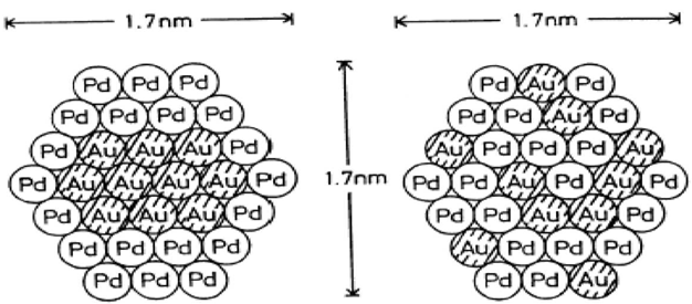
BI-METALLIC NANOPARTICLES, composed of two different metal elements, are of greater interest than monometallic ones, from both the scientific and technological views. They offer a method to control the energy of the Plasmon absorption band of the metallic mixture, which becomes a versatile tool in biosensing. They may also improve the catalytic activity of the particles, sometimes creating new catalysts unknown in the bulk size. Furthermore, structural changes can be created in small bimetallic nanoparticles as a result of alloying of the component metals.
Binary phase diagrams for bulk metals are a well established, commonly utilized tool. However, it is doubtful that they can be extended to the nanometer size regime because of the presence of the large bimetallic interface, the large surface area, and the possible presence of defects at the interface.
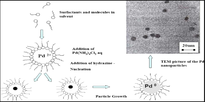
Preparation of Bimetallic Nanoparticles:
Ligands or polymers, especially solvent-soluble polymers, either natural or synthetic, with some affinity for metals are often used as stabilizers of metal nanoparticles. These substances also control both the reduction rate of metal ions and the aggregation process of metal atoms.
STRUCTURE OF BIMETALLIC NANOPARTICLES:
· In bulk metals, atoms are arranged in various geometries, each metal having its own mode of atom placement. The resulting crystal structure is usually simple and depends on the identity of the metal and other conditions such as temperature.
· Bimetallic nanoparticles can adopt another type of structure, in which the distribution of each metal element is not that found in the bulk.
Core-shell structure:
In a core-shell structure, one metal element forms an inner core and the other element surrounds the core to form a shell.

Cross-section and three-dimensional pictures of PVP stabilized Pt core - Pd shell model
Cluster-in-cluster structure:
· In the bimetallic nanoparticles with cluster-in-cluster structures, one element forms nanoclusters and the other element surrounds the nanoclusters and acts as a binder.
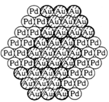
Cluster -in- cluster model of PVP-stabilized Au-Pd (1:1) bimetallic
Alloy Structure:
· In bulk metals, two kinds of metal elements often provide an alloy structure. If the atomic sizes of two elements are similar to each other, then it will be a random alloy.
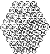
Random model of PVP-stabilized Au-Pd (1:1) bimetallic
PREPARATION OF BIMETALLIC NANOPARTICLES:
1. Co-reduction
2. Successive Reduction
3. Reduction of Double Complexes 4. Electrolysis of Bulk of Metal
Co-reduction of mixed ions:
· Au-Pt bimetallic nanoparticles were prepared by citrate reduction from the corresponding two metal salts, such as
· (III) acid and hexachloroplatinic (IV) acid.
· Polymer-stabilized bimetallic nanoparticles in water-alcohol have been prepared by simultaneous reduction of the two corresponding metal ions with refluxing alcohol.
· For example, colloidal dispersions of Pd-Pt bimetallic nanoparticles can be prepared by refluxing the alcohol-water (1 : 1 v/v) mixed solution of palladium(II) chloride and hexachloroplatinic(IV) acid in the presence of poly(N-vinyl-2-pyrrolidone) (PVP) at about 90-95 °C for 1 h.
· The resulting brownish colloidal dispersions are stable and neither precipitates nor flocculates over a period of several years.
Successive reduction of metal ions:
· Successive reduction of two metal salts can be considered as one of the most suitable methods to prepare core-shell structured bimetallic particles.
· The deposition of one metal element on pre-formed monometallic
nanoparticles of another metal seems to be very effective.
· For this purpose, however, the second element must be deposited on the surface of pre-formed particles, and the pre-formed monometallic nanoparticles must be chemically surrounded by the deposited element.
Reduction of Double Complexes:
· Colloidal silver-platinum alloys have been prepared by NaBH4 reduction of silver (1) bis (oxalato) platinate (II) (Ag2 [Pt (C2O4)2]) in ethylene glycol
Electrochemical Formation of Bimetallic Nanoparticles:
· The Pd-Pt, Ni-Pd, Fe-Co and Fe-Ni bimetallic nanoparticles could be also obtained by this method.
CHARACTERIZATION OF BIMETALLIC NANOPARTICLES:
· The first question asked about metal nanoparticles is concerned with aggregation state, size and morphology.
· Many techniques have been used to reveal the size and homogeneity of metal nanoparticles obtained by chemical methods. As distinct from small organometallic molecules, the composition of metal nanoparticles cannot be so exactly controlled as to be written down as MnM"n .
· However, the homogeneity of particle size or shape on the atomic scale is quite important to reveal the physical properties of nanosized materials.
TRANSMISSION EMISSION SPECTROSCOPY:
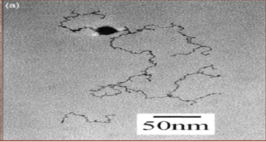
· Metal nanoparticles, especially those consisting of heavy (precious) metal elements, give high contrast when the particles are dispersed on thin carbon films supported by metal grids.
· Sample preparation of colloidal dispersions of metal nanoparticles for TEM observation is quite simple, involving evaporation of a small drop of dispersion onto a carbon-coated micro grid.
· High resolution TEM (HRTEM) can now provide information not only on the particle size and shape but also on the crystallography of monometallic and bimetallic nanoparticles.
NOW YOU CAN ALSO PUBLISH YOUR ARTICLE ONLINE.
SUBMIT YOUR ARTICLE/PROJECT AT articles@pharmatutor.org
Subscribe to PharmaTutor Alerts by Email
FIND OUT MORE ARTICLES AT OUR DATABASE
UV-VIS SPECTROSCOPY:
· The property most immediately observable for metal nanoparticle dispersions of certain metals is their color.
· Comparison of spectra of bimetallic nanoparticles with the spectra of physical mixtures of the respective monometallic particle dispersions can confirm a bimetallic structure for the nanoparticles.
· The absorption peaks that are found in the spectra of Pt and Pd ionic precursors completely disappear after refluxing the alcohol solution of mixed metal ions in the presence of PVP, showing the completion of reduction of both ions.
· Pd-Pt bimetallic nanoparticle dispersions show similar spectra as that of Pd monometallic dispersion in the region of 500-850 nm, but at shorter wavelengths (250-450 nm) the absorbance of Pd-Pt bimetallic nanoparticles is larger than Pd.
· In this wavelength region, the larger the Pt ratio the larger is the absorbance of the bimetallic nanoparticle dispersions. The spectra of bimetallic nanoparticles dispersions were found not only to differ from those of monometallic dispersions, but also from those of physical mixtures. These results suggest the formation of bimetallic nanoparticles.
IR SPECTROSCOPY
· Infrared spectroscopy has been widely applied to the investigation of the surface chemistry of adsorbed small molecules.
· Carbon monoxide, which can be easily adsorbed on metals, is frequently studied. The wave number of adsorbed CO changes dramatically with the binding
structure.
· By comparison of IR spectra of CO on a series of bimetallic nanoparticles at various metal compositions, one can elucidate the surface micro-structure of bimetallic nanoparticles.
X-RAY DIFFRACTION (XRD)
· The presence of bimetallic particles as opposed to a mixture of monometallic particles can also be demonstrated by XRD.
· The diffraction pattern of the physical mixtures consists ofoverlapping lines of the two individual monometallic nanoparticles and is clearly different from that of the bimetallic nanoparticles.
· However, when these metals are divided into small nanoparticles consisting of less than hundreds of atoms, the acquisition of structural information may be difficult.
METAL NMR SPECTROSCOPY:
· NMR spectroscopy of metal isotopes is a powerful technique for understanding the electronic environment of metal atoms in metallic particles by virtue of the NMR shifts caused by free electrons.
· 13CNMR and 1H NMR are also quite useful to understand the structure of adsorbed organic molecules onto the surface of metal nanoparticles.
Applications of bimetallic nanoparticles:
1. Hydrogen storage
2. Improved cathode and anode materials for fuel cells
3. Environmental catalysts
4. Automotive catalysts
5. Bone growth promotors
6. Sunscreens
7. Antibacterial wound dressings
8. Fungicides
9. Biolabeling and detection
10. MRI contrast agents
11. Engineering cutting tool bits
12. Chemical sensors
13. Wear resistant or abrasion resistant coatings
14. Nano pigments
15. Magnetic metal writing powders
16. Structural and physical enhancement of polymers and composites
17. Photo catalyst treatments
18. Nanoscale magnetic particles for high density data storage
19. Electronic circuits and Ferro fluids
20. Opto electronic devices such as switches using rare earth metals
21. Chemical mechanical planarization-CMP
22. Dye sensitized solar cells
Conclusion:
The present review is focused on the bimetallic nanoparticles structure, preparation which are composed of two different metal elements. They are of greater interest than monometallic ones,in terms of size, number of particles, form and crystalline structure, tendency to aggregation, surface reactivity, chemical composition, solubilityindividual susceptibility and interaction of particles with biological components and their biological destiny from both the scientific and technological views which they can be also characterized.It is concluded bimetallic nanoparticles offer a method to control the energy of the Plasmon absorption band of the metallic mixture, which becomes a versatile tool in biosensing. They may also improve the catalytic activity of the particlesand these substances also control both the reduction rate of metal ions and the aggregation process of metal atoms. The same concept can be also remains to be explored regarding the potential of the bimetallic nanoparticles but present they are potntially improving catalytic activity of many processes.The development of new materials in this field is being pursued intensively and the health effects cannot all be studied (Won Kang and Hwang, 2004). Moreover, toxicological tests will have to consider that bi metallic nanoparticles surfaces normally are altered to prevent aggregation. They are having numerous applications like Photo induced hydrogen generation from water, Hydrogenation of olefins, Effects of ligands in catalysis, hydration of acrylonitrile etc., besides them bimetallic nano particles shows very interesting magnetic properties.
REFERENCES:
1. V j Mohan raj and y chen, nanoparticles-a review,tjpr journal 2005(5),561-573.
2. Asia Pacific Nanotechnology Forum, 2005, http://www.apnf.org.
3. Afaq F, Abidi P, Matin R and Rahman Q. Cytotoxicity, pro-oxidant effects and antioxidant depletion in rat lung alveolar macrophages exposed to ultrafine titanium dioxide. JAppl Toxicol 1998, 18, 307-312.
4. Aitken RJ, Creely KS and Tran CL. Nanoparticles: An Occupational Hygiene Review First International Symposium on Occupational Health Implications of Nanomaterials. 12-14 October 2004 Palace Hotel, Buxton, Derbyshire, UK
5. http://www.hsl.gov.uk/capabilities/nanosymrep_ffinal.pdf
6. Akerman ME, Chan WCW, Laakkonen P, Bhatia SN and Ruoslahti E. Nonocrystal
7. targeting in vivo. PNAS 2002, 99, 12617-12621.
8. Aprahamian M, Michel C, Humbert W, Devissaguet J.P and Damge C. Transmucosal passage of polyalkylcyanoacrylate nanocapsules as a new drug carried in the small intestine. Biol Cell 1987, 61, 69-76.
9. Aue WP, Bartholdi E and Ernst RR. 2-dimensional spectroscopy - application to nuclear magnetic-resonance. J Chem Phys 1976, 64, 2229-2246.
10. Australian Academy of Sciences. Nanotechnology Benchmark Project, 2005, http://science.org.au/policy/nano-report.pdf.
11. Bain CD, Troughton EB, Tao YT, Evall J, Whitesides GM and Nuzzo RG. Formation of monolayer films by the spontaneous assembly of organic thiols from solution onto gold. JAmer Chem Soc 1989, 111, 321-335.
12. Baran ET, Ozer N and Hasirci V. In vivo half life of nanoencapsulated L-asparaginase. J Mat Sci: Mat in Med 2002, 13, 1113-1121.
13. Bazile D, Prud'Homme C, Bassoullet M-T, Marlard M, Spenlehauer G and Veillard M. Stealth PEG-PLA nanoparticles avoid uptake by the mononuclear phagocytes system. JPharm Sci 1995, 84, 493-498.
14. Berry CC, Wells S, Charles S and Curtis ASG. Dextran and albumin derivatised iron oxide nanoparticles influence on fibroblasts in vitro. Biomaterials 2003, 24, 4551-4557.
15. BIA. Report 7/2003e; BIA-Workshop. Ultrafine aerosols at workplaces. BG Institute for Occupational Safety and Health - BIA, Sankt Augustin, Germany.
16. Binnig G and Rohrer H. Scanning Tunneling Microscopy Helvetica Physica Acta 1982,
17. 55, 726-735.
18. Borm PJ. Particle toxicology: from coal mining to nanotechnology. Inhal Toxicol 2002, 14, 311-324.
19. Borm PJ and Kreyling WG. Toxicological hazards of inhaled nanoparticles - potential implications for drug delivery. J Nanoscience Nanotechnol 2004, 4, 521-531.
20. Bourges JL, Gautier SE, Delie F, Bejjani RA, Jeanny JC, BenEzra D and Behar-Cohen FF. Ocular drug delivery targeting the retina and retinal pigment epithelium using polylactide nanoparticles. Invest Ophthalmol Vis Sci 2003, 44, 3562-3569.
21. BSI, British Standards Institution. Vocabulary - Nanoparticles, Publicly Available Specification, PAS 71:2005. BSI. London.
22. Brown DM, Wilson MR, MacNee W, Stone V and Donaldson K. Size dependent
23. Cassee FR, Muijser H, Duistermaat E, Freijer JJ, Geerse KB, Marijnissen JC and Arts JH. Particle size-dependent total mass deposition in lung determines inhalation toxicity of cadmium chloride aerosols in rats. Application of a multiple path dosimetry model.
24. Arch Toxicol 2002, 76, 277-286.
25. Chan WCW and Nie SM. Quantum dot bioconjugates for ultrasensitive nonisotopic detection. Science 1998, 281 (5385), 2016-2018.
26. Clark J, Singer EM, Korns DR and Smith SS. Design and analysis of nanoscale bioassemblies. Biotechniques 2004, 36, 992.
27. Colvin VL. The potential environmental impact of engineered nanoparticles, Nature Biotechnology 2003, 21, 1166-1170.
28. Cruz T, Gaspar R, Donato A and Lopes C. Interaction between polyalkylcyanoacrylate nanoparticles and peritoneal macrophages: MTT metabolism, NBT reduction and NO production. Pharm Res 1997, 14, 73-79.
29. Cui Z and Mumper RJ. Topical immunization using nanoengineered genetic vaccines. J ControlledRel. 2002, 81, 173-184.
30. Cui Z and Mumper RJ. Microparticles and nanoparticles as delivery systems for DNA vaccines. Crit Rev Ther Drug Carrier Syst 2003, 20, 103-137.
31. W.H. de Jong, B. Roszek, R.E. Geertsma: Nanotechnology in medical applications: Possible risks for human health, RIVM report 265001002/2005
32. Dick CA, Brown DM, Donaldson K and Stone V. The role of free radicals in the toxic and inflammatory effects of four different ultrafine particle types. Inhal Toxicol 2003, 15,
33. 39-52.
34. Donaldson K, Stone V, Tran CL, Kreyling W and Borm PJA. Nanotoxicology. Occup
35. Environ Med 2004, 61, 727-728.
36. Emerich DF and Thanos CG. Nanotechnology and medicine. Expert Opinion Biol
37. Therapy 2003, 3, 655-663.
38. Ferrari M. Cancer nanotechnology: opportunities and challenges. Nature Reviews:
39. Cancer 2005, 5, 161-171.
40. Frens G. Controlled nucleation for regulation of particle-size in monodisperse gold suspensions. Nature -Physical Science 1973, 241, 20-22.
41. Gemeinhart RA, Luo D and Saltzman WM. Cellular fate of modular DNA delivery system mediated by silica nanoparticles. Biotechn Prog 2005, 21, 532-537.
NOW YOU CAN ALSO PUBLISH YOUR ARTICLE ONLINE.
SUBMIT YOUR ARTICLE/PROJECT AT articles@pharmatutor.org
Subscribe to PharmaTutor Alerts by Email
FIND OUT MORE ARTICLES AT OUR DATABASE


